Invasive ductal carcinoma in a 51-year-old male: case report
SPMC J Health Care Serv. 2019;5(2):6 ARK: http://n2t.net/ark:/76951/jhcs6ct44a
1Department of General Surgery, Southern Philippines Medical Center, JP Laurel Ave, Davao City, Philippines
Correspondence Stephen Matthew B Santos, stephensantos4@gmail.com
Article editor Wesley Reyes
Received 19 October 2018
Accepted 4 November 2019
Cite as Santos SMB, Borje EA. Invasive ductal carcinoma in a 51-year-old male: case report. SPMC J Health Care Serv. 2019;5(2):6. http://n2t.net/ark:/76951/jhcs6ct44a
Introduction
Male breast cancer (MBC) constitutes only 1% of all cases of breast cancer worldwide.1 2 3 4 In 2014, MBC had an estimated global incidence of 8,000 cases according to the World Health Organization.5 Two types of breast cancer are seen in males, the most common type of which is invasive ductal carcinoma (IDC).6 The other type, invasive lobular carcinoma is less commonly seen due to the absence of lobules in male breasts.7 MBC is usually associated with risk factors such as genetic mutations in the BRCA1 and BRCA2 genes, and exposure to ionizing radiation or endocrine-disrupting compounds.8 9 10 Other conditions, such as Klinefelter’s syndrome, obesity, and hyperestrogenism, are also associated with MBC.11 Among patients with MBC, the median age at diagnosis is between 62 and 67 years.1 2 8 12 Patients with MBC usually present with a palpable subareolar mass or swelling and may have symptoms such as nipple involvement with ulceration or bleeding, gynecomastia and axillary lymphadenopathy.1 Unlike in female breast cancer (FBC), histological grading in MBC has no prognostic value.13 14 For MBC, the surgical treatment of choice is usually a modified radical mastectomy,15 while for FBC, women may opt for a breast conservative surgery, which is less invasive and has better cosmetic outcomes.16 We present the case of a 51-year-old male who came in due to an enlarging right breast mass, an IDC, for which he subsequently underwent a modified radical mastectomy.
Clinical features
A 51-year-old male consulted in our department complaining of a gradually enlarging right-sided breast mass. In the past seven years, the patient has had a recurring subareolar nodule in the right breast with secretions ranging from clear to white, to bloody nipple discharge. The nodule was surgically removed twice—six years ago and a year ago—but the histological diagnosis was unknown each time. The patient reported that the breast mass was not completely removed during the previous surgeries. The current breast mass, for which the patient consulted, slowly increased in size within the last six months prior to consultation. It also became painful a few weeks before the patient came to our outpatient clinic.
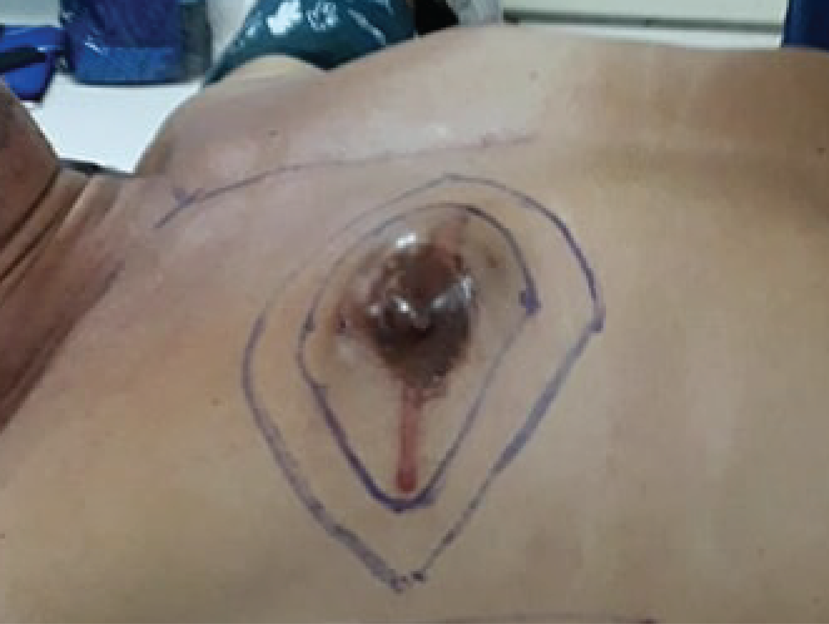
The patient has been a smoker for three pack-years and a chronic alcoholic beverage drinker consuming hard liquor everyday for 30 years. He had no known comorbidities, family history of cancer, or exposure to environmental risk factors. He denied other accompanying signs or symptoms such as fever, weight loss, nipple retraction, or skin dimpling on the breast. On physical examination, the patient had a 5-cm-long hypertrophic scar above the right nipple. A 4 cm x 5 cm x 1 cm nontender mass could be palpated beneath the hypertrophic scar. The whole right nipple was surrounded by a smooth, thinning skin (Figure 1).
|
|
Figure 1 Right breast mass measuring 4 x 5 x 1 cm. |
There were no palpable axillary or cervical lymphadenopathies. The contralateral chest and axilla were normal, and there was no gynecomastia. The patient had normal-looking external genitalia with palpable bilateral testes. The rest of the physical examination findings were unremarkable.
Diagnostic approaches
Complete blood count, electrolytes, creatinine, and liver function tests on admission were all normal. The liver ultrasound and the chest radiograph were also unremarkable. Breast ultrasonography showed a 6.6 cm x 4.3 cm x 5.6 cm circumscribed, complex mass with hypoechoic, anechoic and mobile dependent low-level echoes on the right chest. The surrounding right breast tissues and the left breast appeared normal.
A core needle biopsy of the right breast mass showed sections of glandular structures lined by atypical cells with hyperchromatic nuclei. Some of the cells were arranged in cohesive sheets and haphazardly invaded the surrounding connective tissues. These histopathological findings are consistent with IDC. We proceeded to manage the patient breast cancer, Stage IIA (cT2N0M0).
Therapeutic approaches
The patient underwent a right-sided modified radical mastectomy (MRM). We made an incision around the lesion for the formation of upper and lower flaps and elevated the breast from the pectoralis fascia. A 4x5x1 cm mass was noted at the subareolar area, with no involvement of the pectoralis major muscle. On axillary dissection, we removed the fibrofatty tissues of the right axilla, and the level-I and level-II axillary lymph nodes (Figure 2).

|
|
Figure 2 The right pectoralis major muscle, exposed right after modified radical mastectomy. |
Grossly, the excised specimen was composed of the right nipple, areola, subareolar tumor (pT2), surrounding skin, subcutaneous fat, and axillary lymph nodes (Figure 3). Histological examination of the breast tissue showed presence of atypical cells, the majority of which are forming glandular structures with moderate tubule formation of 10% to 75%. These atypical cells exhibit nuclear hyperchromatism with moderate pleomorphism (Figure 4). Tumor cells were found in two out of 17 harvested lymph nodes, blood vessels, and lymphatic vessels. However, all margins of resection were negative for tumor cells. The final histopathologic diagnosis was IDC (Nottingham grade 1).

|
|
Figure 3 The excised breast specimen showing the subareolar tumor and axillary lymph nodes. |

|
|
Figure 4 Histopathology of the right breast mass at low power field (A: hematoxylin-eosin stain, x10) and high power field (B: hematoxylin-eosin stain, x40) showing atypical cells forming glandular structures (A: yellow ring) and exhibiting nuclear hyperchromatism (B: green arrows) with moderate pleomorphism (B: yellow arrows). |
The patient's postoperative course was unremarkable, and we discharged him from the hospital on the third postoperative day.
Subsequent immunohistochemistry tests on the specimens were positive for estrogen receptor (ER) and progesterone receptor (PR), but negative for HER2/neu. The patient’s final diagnosis after the postoperative biopsy result was: IDC, stage IIB (pT2N1M0), ER/PR-positive.
The patient was started on adjuvant chemotherapy 3 weeks after surgery using doxorubicin and cyclophosphamide regimen, given every 21 days for a total of four cycles . After the fourth cycle, the patient also started receiving paclitaxel every 14 days for a total of four cycles. Two weeks after the last cycle of paclitaxel, we started the patient on tamoxifen 20 mg per day.
Outcomes
We did not observe any adverse chemotherapeutic drug reactions, tumor recurrence, or appearance of new tumors in the contralateral breast. Ultrasound of the contralateral breast done five months after the last cycle of adjuvant chemotherapy, one year after surgery, revealed normal results. As of this writing, the patient continues to take daily oral tamoxifen. For two years now, the patient regularly returns for follow-up checkup every three months at at our cancer clinic. As of his last visit, the patient had no signs of recurrence on the post-operative site (Figure 5), and had a normal chest radiograph and liver ultrasound findings. We also advised the patient to undergo complete physical examination twice yearly and mammography annually to detect breast cancer recurrence.

|
|
Figure 5 Postmastectomy scar 21 months after surgery. |
Discussion
Our patient's recurring breast mass was removed twice in the past eight years without a definitive diagnosis. This precluded an early therapeutic intervention for what turned out to be an IDC.
Breast mass and nipple discharge are symptoms that, when found in men, are highly suggestive of MBC.17 Breast cancer in males and females are relatively similar in terms of etiology, diagnosis, and treatment.18 However, MBC is rare and information regarding this condition is limited, making early diagnosis and intervention uncommon.
The more common types of MBC are IDC and ductal carcinoma in situ (DCIS).6 Infiltrating lobular carcinoma, a malignancy in the milk-producing glands, occurs less commonly in males. While the female breast is predominantly composed of lobules that produce milk, ducts, glandular epithelium, and non-adipose stroma, the normal male breast has no lobules, and is composed mostly of adipose tissue with few ducts and periductal stroma.19
MBC presenting as DCIS comprise 10% of all MBCs. Of all DCIS cases, only 5% are pure DCIS, while the rest eventually develop into IDC in a span of a decade or more.20 21 Incompletely excised lesions of low-grade DCIS have the proclivity for local recurrence, with possible invasion at a protracted time course. On the other hand, high-grade DCIS that is left untreated or inadequately treated will evolve into an IDC in less than 5 years.22 Our patient may have initially had DCIS eight years ago. Excision of the DCIS may not have been thorough enough as to achieve fully negative margins, and through time, the DCIS progressed into an IDC. Studies in the past years highly emphasize the role of health care in promoting public awareness about MBC in order to facilitate early recognition and treatment of the disease.23
The main risk factors of MBC are Klinefelter’s syndrome and a positive family history of breast or ovarian malignancy.1 2 3 However, genetic predisposition is also considered to be a major risk factor of MBC.24 Male patients with a positive family history of breast cancer are most likely carriers of BRCA2 gene mutations, and are likely to be offered genetic counseling and testing.25 The risk of having MBC at age 70 years is 6% for male patients who have BRCA2 mutations.26 Other risk factors include the use of external estrogen or testosterone, and a history of orchitis or epididymitis diagnosed at 50 years or older.2 Our patient, however, had none of these risk factors.
The definitive diagnosis of MBC is largely made through core needle biopsy and cytology. MBC tends to be localized in the subareolar region,27 as in our patient, and can be identified by mammography and/or ultrasonography.28 In mammography, malignant microcalcifications are less commonly seen in MBC than in female breast cancer (FBC).29
MBC occurs later in life, with a median age at diagnosis of 67 years,30 and is usually diagnosed at an advanced clinical stage.31 Breast cancer in men are typically low-grade, and usually estrogen- and progesterone-receptor positive, in contrast to breast cancer in women, which can be aggressive and are frequently triple-negative or HER2-positive.13 31 32 When positive for HER2 receptor, MBC would indicate a worse prognosis for the patient.33 Our patient was diagnosed with stage IIB ER/PR-positive, HER2-negative breast cancer eight years after the onset of symptoms, which may indicate a low-grade disease diagnosed at an advanced stage.
Due to the rarity of MBC, management strategies for the condition are extrapolated from randomized clinical studies in women. Current surgical treatment options for MBC include modified radical mastectomy (approximately 70% of all cases), followed by radical mastectomy (8 to 30%), simple or total mastectomy (5 to 14%), and lumpectomy with or without radiation (1 to 13%).15 34 We did a modified radical mastectomy with axillary lymph node dissection on our patient. Although some surgeons would opt for a breast conservative surgery (BCS) such as lumpectomy, quadrantectomy, partial mastectomy or segmental mastectomy, modified radical mastectomy with sentinel lymph node biopsy or axillary lymph node dissection still remains the gold standard of surgical treatment of MBC.35 BCS is less used in MBC since most lesions are located in the subareolar region, usually with nipple involvement, requiring a more extensive surgery such as mastectomy.27 36 Also, the small size of male breast makes it difficult to achieve negative or tumor-free margins when BCS is performed.30
The ER/PR-positive, HER2-negative results of our patient on the immunohistochemistry test, and the presence of lymph node involvement on the postoperative biopsy results, prompted the decision to give hormone therapy (tamoxifen) as adjuvant treatment, together with dose-dense adjuvant chemotherapy of doxorubicin-cyclophosphamide and paclitaxel combination. Hormone therapy can be used as first-line treatment for hormone-receptor positive MBC,30 and as adjuvant or palliative therapy for advanced cases.20 The most widely used systemic hormonal treatment for MBC in adjuvant and metastatic settings is tamoxifen, a selective estrogen receptor modulator usually taken for 5 years and frequently associated with significant adverse effects.28 Aromatase inhibitors (AI), a new treatment option used more commonly in postmenopausal hormone-sensitive FBC, have also gained popularity in the treatment of MBC. Current studies do not favor their use in early disease.37
In combination with hormonal treatment, dose-dense adjuvant cytotoxic chemotherapy with an anthracycline- and taxane-based regimen, may prove beneficial in patients with lymph node involvement, tumors larger than 1 cm, patients in high-risk groups, and in younger patients.2 28 The use of adjuvant chemotherapy has been shown to reduce cancer recurrence and improve overall survival.36 Radiation therapy is considered as adjuvant treatment for patients with T3 or higher tumors, four or more positive lymph nodes, and positive surgical margins.38 In combination with BCS, radiation therapy may also be used to decrease local tumor recurrence,39 but its usefulness is limited due to its tendency to cause cardiovascular and pulmonary complications in older patients.37
Although the pathophysiology of MBC is relatively similar to that of post-menopausal FBC,2 variations in their medical management—especially in the treatment of ER-positive cancers—have been identified based on the biological differences between males and females.20 In postmenopausal FBC, aromatase inhibitors have been found to be more effective compared to tamoxifen, which is frequently used in premenopausal FBC.40 Postmenopausal women produce estrogen in the peripheral tissues of the body (i.e., liver, fat, muscle, skin, and breast), while premenopausal women produce estrogen mainly in the ovaries. AI are not used in premenopausal women since high circulating levels of androgen in this population compete with AI in the aromatase enzyme complex, thus rendering the AI ineffective. Also, the hypothalamic-pituitary feedback mechanism that detects low levels of serum estrogen in premenopausal women, upregulates the production of aromatase enzyme in the ovary, which would counteract the effect of AI in this population.41 In the case of MBC, the administration of AI is associated with significant increases in FSH, LH, and testosterone levels, but with no change in estradiol levels.42 43 Consequently, high testosterone levels may saturate the aromatase enzyme complex, leaving no free aromatase for the AI to act on.44 45 Additionally, tamoxifen has been favored as first-line adjuvant treatment because aromatase inhibitors are associated with severe complications, e.g., bone loss and toxic cardiopulmonary effects, among men. A 1.5-fold increase in mortality had been observed in male patients treated with AI compared to those receiving tamoxifen for MBC.20
MBC in younger males tend to have poor prognosis because most cases are dismissed as gynecomastia.8 Males have poorer prognosis than females, since most MBC cases are diagnosed late.46 FBC histological grading determines prognosis, such that a higher-grade cancer corresponds to a significantly reduced overall survival.13 32 In contrast, MBC histological grading has no significant correlation with breast cancer outcomes. Several factors, such as low mitotic activity index, presence of fibrotic focus, high density of tumour-infiltrating lymphocytes, luminal HER2/neu positive subtype, and absence of lymphovascular invasion all strongly correlate with survival. 13
The strongest predictor for both local recurrence and metastasis in MBC is axillary nodal involvement.28 The risk for contralateral breast cancer is relatively high in males who are initially diagnosed before the age of 50 years. Thus, periodic screening is imperative for these patients.46 For our 51-year-old patient with axillary nodal involvement noted after mastectomy, ultrasonography one year after surgery is necessary to check for recurrence and appearance of contralateral breast cancer. There are no specific clinical practice guidelines for MBC treatment and follow-up, so most of the management approaches for MBC are usually based on guidelines for FBC. Annual mammography for tumor recurrence or second breast cancers and screening for non-breast second malignancies—such as prostate, lung, colorectal, and esophageal cancers—are recommended for optimal surveillance. MBC survivors should also undergo twice yearly physical examination for the first 5 years after surgery, and then annually thereafter.34 47
In summary, we were presented with a male patient complaining of an eight-year history of unilateral breast mass. The mass was removed twice, each time without a definitive diagnosis. He subsequently underwent a modified radical mastectomy, cytotoxic adjuvant chemotherapy consisting of a dose-dependent doxorubicin-cyclophosphamide and paclitaxel combination, followed by hormone therapy with tamoxifen. Due to the rarity of MBC and its worse prognosis compared to FBC, a high index of suspicion is required among practitioners who encounter any breast mass among males. Increasing the level of public awareness on MBC can prevent delays in the diagnosis and treatment of the disease.
Contributors
SMBS and EAB both contributed to the diagnostic and therapeutic care of the patient in this report. Both of them acquired relevant patient data, and searched for and reviewed relevant medical literature used in this report. Both wrote the original draft, performed the subsequent revisions, approved the final version, and agreed to be accountable for all aspects of this report.
Patient consent
Obtained
Reporting guideline used
CARE Checklist
(https://www.care-statement.org/checklist)
Article source
Submitted
Peer review
External
Competing interests
None declared
Access and license
This is an Open Access article licensed under the Creative Commons Attribution-NonCommercial 4.0 International License, which allows others to share and adapt the work, provided that derivative works bear appropriate citation to this original work and are not used for commercial purposes. To view a copy of this license, visit http://creativecommons.org/licenses/by-nc/4.0/
References
1. Sanguinetti A, Polistena A, Lucchini R, Monacelli M, Galasse S, Avenia S, Triola R, Bugiantella W, Cirocchi R, Rondelli F, Avenia N. Male breast cancer, clinical presentation, diagnosis and treatment: Twenty years of experience in our Breast Unit. Int J Surg Case Rep. 2016;20S(Suppl):8-11.
2. Yalaza M, İnan A, Bozer M. Male Breast Cancer. J Breast Health. 2016 Jan 1;12(1):1-8.
3. Fiala L, Coufal O, Fait V, Foretová L. [Male breast cancer--our experience]. Rozhl Chir. 2010 Oct;89(10):612-8.
4. Onami S, Ozaki M, Mortimer JE, Pal SK. Male breast cancer: an update in diagnosis, treatment and molecular profiling. Maturitas. 2010 Apr;65(4):308-14.
5. Shapiro CL. Comparing male and female breast cancer. 2017 Aug 14 [cited 2019 Dec 17]. In: CURE - cancer updates, research and education [Internet]. New Jersey: CURE Media Group. C2019. Available from: https://www.curetoday.com/publications/cure/2017/rare-cancer-summer-2017/comparing-male-and-female-breast-cancer.
6. Memorial Sloan Kettering Cancer Center. Types of male breast cancer [Internet]. New York: Memorial Sloan Kettering Cancer Center. C2019. Available from: https://www.mskcc.org/cancer-care/types/breast-male/types.
7. Barry S, Ha KY, Laurie L. Carcinoma of the breast in men. Proc (Bayl Univ Med Cent). 2012 Oct;25(4):367-8.
8. Madeira M, Mattar A, Passos RJ, Mora CD, Mamede LH, Kishino VH, Torres TZ, de Sá AF, dos Santos RE, Gebrim LH. A case report of male breast cancer in a very young patient: what is changing? World J Surg Oncol. 2011 Feb 3;9:16.
9. Pritzlaff M, Summerour P, McFarland R, Li S, Reineke P, Dolinsky JS, et al. Male breast cancer in a multi-gene panel testing cohort: insights and unexpected results. Breast Cancer Res Treat. 2017 Feb;161(3):575-586.
10. Fenga C. Occupational exposure and risk of breast cancer. Biomed Rep. 2016 Mar;4(3):282-292.
11. Ruddy KJ, Winer EP. Male breast cancer: risk factors, biology, diagnosis, treatment, and survivorship. Ann Oncol. 2013 Jun; 24(6):1434-43.
12. André S, Pereira T, Silva F, Machado P, Vaz F, Aparício M, et al. Male breast cancer: Specific biological characteristics and survival in a Portuguese cohort. Mol Clin Oncol. 2019 Jun;10(6):644-654.
13. Vermeulen MA, Slaets L, Cardoso F, Giordano SH, Tryfonidis K, van Diest PJ, et al. Pathological characterisation of male breast cancer: Results of the EORTC 10085/TBCRC/BIG/NABCG International Male Breast Cancer Program. Eur J Cancer. 2017 Sep;82:219-227.
14. Schwartz AM, Henson DE, Chen D, Rajamarthandan S. Histologic grade remains a prognostic factor for breast cancer regardless of the number of positive lymph nodes and tumor size: a study of 161,708 cases of breast cancer from the SEER Program. Arch Pathol Lab Med. 2014 Aug;138(8):1048-52.
15. Sousa B, Moser E, Cardoso F. An update on male breast cancer and future directions for research and treatment. Eur J Pharmacol. 2013 Oct 5;717(1-3):71-83.
16. McCrate F, Dicks E, Powell E, Chafe J, Roome R, Simmonds C, Etchegary H. Surgical treatment choices for breast cancer in Newfoundland and Labrador: a retrospective cohort study. Can J Surg. 2018 Dec 1;61(6):377-384.
17. Farooq A, Horgan K. Male breast cancer presenting as nipple discharge. Case Reports in Surgery. 2011; Volume 2011, Article ID 804843, 3 pages.
18. Grimm LJ. Male breast cancer imaging. Medscape. Updated April 11, 2019.
19. Iuanow E, Kettler M, Slanetz PJ. Spectrum of disease in the male breast. AJR Am J Roentgenol. 2011;196:W247-W259.
20. Fentiman IS. Male breast cancer is not congruent with the female disease. Crit Rev Oncol Hematol. 2016 May;101:119-24.
21. Erbas B, Provenzano E, Armes J, Gertig D. The natural history of ductal carcinoma in situ of the breast: a review. Breast Cancer Res Treat. 2006 May;97(2):135-44.
22. Sanders ME, Schuyler PA, Simpson JF, Page DL, Dupont WD. Continued observation of the natural history of low-grade ductal carcinoma in situ reaffirms proclivity for local recurrence even after more than 30 years of follow up. Mod Pathol. 2015;28:662–9.
23. White J, Kearins O, Dodwell D, Horgan K, Hanby AM, Speirs V. Male breast carcinoma: increased awareness needed. Breast Cancer Res. 2011;13(5):219. Published 2011 Sep 29.
24. Katz J. Breast cancer risk factors. Medscape. Updated December 16, 2019.
25. Wolpert N, Warner E, Seminsky MF, Futreal A, Narod SA. Prevalence of BRCA1 and BRCA2 mutations in male breast cancer patients in Canada. Clin Breast Cancer. 2000 Apr;1(1):57-63; discussion 64-5.
26. Tai YC, Domchek S, Parmigiani G, Chen S. Breast cancer risk among male BRCA1 and BRCA2 mutation carriers. J Natl Cancer Inst. 2007 Dec 5;99(23):1811-14.
27. Doyle S, Steel J, Porter G. Imaging male breast cancer. Clin Radiol. 2011 Nov;66(11):1079-85.
28. Korde LA, Zujewski JA, Kamin L, Giordano S, Domchek S, Anderson WF, Bartlett JM, Gelmon K, Nahleh Z, Bergh J, Cutuli B, Pruneri G, McCaskill-Stevens W, Gralow J, Hortobagyi G, Cardoso F. Multidisciplinary meeting on male breast cancer: summary and research recommendations. J Clin Oncol. 2010 Apr 20;28(12):2114-22.
29. Chen L, Chantra PK, Larsen LH, Barton P, Rohitopakarn M, Zhu EQ, Bassett LW. Imaging characteristics of malignant lesions of the male breast. RadioGraphics. 2006; 26(4):993-1007.
30. Giordano SH, Cohen DS, Buzdar AU, Perkins G, Hortobagyi GN. Breast carcinoma in men: a population-based study. Cancer. 2004 Jul 1;101(1):51-7.
31. Anderson WF, Jatoi I, Tse J, Rosenberg PS. Male breast cancer: a population-based comparison with female breast cancer. J Clin Oncol. 2010 Jan 10;28(2):232-9.
32. Breastcancer.org. Breast cancer in men biologically different than breast cancer in women [Internet]. Pennsylvania: Breastcancer.org. C2019 [cited 2019 Dec 17]. Available from: https://www.breastcancer.org/research-news/male-bc-differs-biologically-from-female.
33. Bruce DM, Heys SD, Payne S, Miller ID, Eremin O. Male breast cancer: clinico-pathological features, immunocytochemical characteristics and prognosis. Eur J Surg Oncol. 1996 Feb;22(1):42-6.
34. Cutuli B. Strategies in treating male breast cancer. Expert Opin Pharmacother. 2007 Feb;8(2):193-202.
35. Giunta G, Rossi M, Toia F, Rinaldi G, Cordova A. Male breast cancer: Modified radical mastectomy or breast conservation surgery? A case report and review of the literature. Int J Surg Case Rep. 2017;30:89-92.
36. American Cancer Society. Surgery for breast cancer in men [Internet]. Atlanta: American Cancer Society. C2019 [cited 2019 Dec 17]. Available from: https://www.cancer.org/cancer/breast-cancer-in-men/treating/surgery.html.
37. Patten DK, Sharifi LK, Fazel M. New approaches in the management of male breast cancer. Clin Breast Cancer. 2013 Oct;13(5):309-14.
38. Jana BRP. Breast cancer in men overview of male breast cancer. 2019 Oct 9 [cited 2019 Dec 17]. In: Medscape. New York: Medscape. C2019. Available from: https://emedicine.medscape.com/article/1954174-overview#a4.
39. Pant K, Dutta U. Understanding and management of male breast cancer: a critical review. Med Oncol. 2008;25(3):294-298.
40. Untch M, Jackisch C. Exemestane in early breast cancer: a review. Ther Clin Risk Manag. 2008 Dec;4(6):1295-304. doi: 10.2147/tcrm.s4007. PMID: 19337436; PMCID: PMC2643110.
41. Burstein H. Aromatase Inhibitors Vs Tamoxifen: A Changing of the Guard? - Medscape - Feb 14, 2002.
42. Zagouri F, Sergentanis TN, Koutoulidis V, Sparber C, Steger GG, Dubsky P, et al. Aromatase inhibitors with or without gonadotropin-releasing hormone analogue in metastatic male breast cancer: a case series. Br J Cancer. 2013 Jun 11;108(11):2259-63.
43. Bighin C, Lunardi G, Del Mastro L, Marroni P, Taveggia P, Levaggi A, Giraudi S, Pronzato P. Estrone sulphate, FSH, and testosterone levels in two male breast cancer patients treated with aromatase inhibitors. Oncologist. 2010;15(12):1270-2. doi: 10.1634/theoncologist.2010-0102. Epub 2010 Dec 8. PMID: 21147874; PMCID: PMC3227929.
44. Eggemann H, Ignatov A, Smith BJ, Altmann U, Von Minckwitz G, Rohl FW, Jahn M, Costa SD. Adjuvant therapy with tamoxifen compared to aromatase inhibitors for 257 male breast cancer patients. Breast Cancer Res Treat. 2013; 137, 465–470.
45. Rachid S, Yacouba H, Hassane N. Male breast cancer: 22 case reports at the National Hospital of Niamey-Niger (West Africa). Pan Afr Med J. 2009 Nov 16;3:15.
46. Rachid S, Yacouba H, Hassane N. Male breast cancer: 22 case reports at the National Hospital of Niamey-Niger (West Africa). Pan Afr Med J. 2009 Nov 16;3:15.
47. Ferzoco RM, Ruddy KJ. Optimal delivery of male breast cancer follow-up care: improving outcomes. Breast Cancer (Dove Med Press). 2015 Nov 23;7:371-9. doi: 10.2147/BCTT.S75630. PMID: 26648754; PMCID: PMC4664432.
Copyright © 2019 SMB Santos, et al.

Published
December 31, 2019
Issue
Volume 5 Issue 2 (2019)
Section
Case report

This work is licensed under a Creative Commons Attribution-NonCommercial 4.0 International License.
Authors who publish with this journal agree to the following terms:
- Authors retain copyright and grant the journal right of first publication with the work simultaneously licensed under a Creative Commons Attribution-NonCommercial 4.0 International License that allows others to share the work for non-commercial purposes with an acknowledgement of the work's authorship and initial publication in this journal.
- Authors are able to enter into separate, additional, non-commercial contractual arrangements for the non-exclusive distribution of the journal's published version of the work (e.g., post it to an institutional repository or publish it in a book), with an acknowledgement of its initial publication in this journal.
- Authors grant the journal permission to rewrite, edit, modify, store and/or publish the submission in any medium or format a version or abstract forming part thereof, all associated supplemental materials, and subsequent errata, if necessary, in a publicly available publication or database.
- Authors warrant that the submission is original with the authors and does not infringe or transfer any copyright or violate any other right of any third parties.